The Tale of Typhoid Mary

As mentioned previously, Typhoid Fever  which is caused by Salmonella typhi was thought to be a sickness affecting only unprivileged communities. That is why when the sickness began reaching well-off families of New York and Long Island, homeowners hired the Department of Health to investigate.

After some exploration into the households effected, a common link was found. Each family had fallen ill about 3 weeks after hiring Mary Mallon, an immigrant from Ireland, as their cook or domestic servant. By the year 1907 when the

DOH caught up with Mary, they had connected her with 22 cases of Typhoid Fever. They concluded that the was a healthy carrier of the bacteria, infecting majority of those she cooked for.

When confronted, Mary was said to have chased the inspector away with a rolling-pin. The police were involved yet she managed to elude capture for another 5 hours. Not due to guilt, more so due to the fact that she did not understand. Eventually she was submitted to a stool test and the results came back positive for S. typhi. Treatments were attempted, and multiple tests were done while Mary was quarantined against her will for a period of three years. Eventually Mary was released on the condition that she would never cook again.  After being released, Mary Mallon changed her name to Mary Brown and began cooking again, eluding authorities for 5 years. Many more people were ailed and two more people died.

In many articles Mary comes across as a callous and knowingly contagious carrier. However many sources say that she would stay with the family and care for them through the illness. It has also been established that Mary was never properly educated on her situation and remained in denial of her condition her entire life. In addition to her lack of education, Mary was an immigrant and cooking was her trade. Although she was offered assistance in finding a non cooking job, it is said that she never understood why she caused a threat to others, so she of course went back to doing what she enjoyed and what she was good at.

Mary was recaptured and it was later found that she had  spread the disease directly to 51 people causing 3 deaths. Indirectly, it is estimated that approximately 400 people were exposed to the disease due to those infected by Typhoid. Mary Mallon lived out her days alone and died a slow death after a total of 26 years in enforced isolation.


Brooks J. The sad and tragic life of Typhoid Mary. CMAJ: Canadian Medical Association Journal. 1996;154(6):915-916.

Marineli F, Tsoucalas G, Karamanou M, Androutsos G. Mary Mallon (1869-1938) and the history of typhoid fever. Annals of Gastroenterology : Quarterly Publication of the Hellenic Society of Gastroenterology. 2013;26(2):132-134.

The Editors of Encyclopædia Britannica. (2017). Typhoid Mary. Encyclopaedia Britannica. Retrieved from https://www.britannica.com/biography/Typhoid-Mary. Accessed 12/05/2017.

Photo Sources:
Who is more at risk?

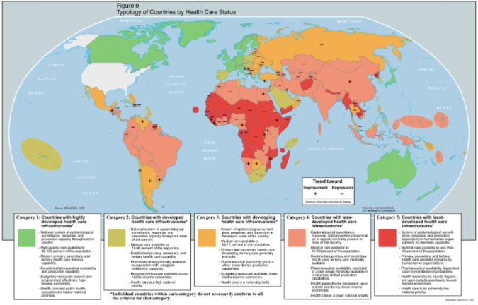
Poverty and M.tuberculosis are closely linked with each other. The main causative agent for tuberculosis is M.tuberculosis. Tuberculosis is ranks alongside HIV/AIDS as the top notch infectious killer worldwide with 9.6 million of new incident cases and 1.5 million death estimated to have been occurred in 2014[7]. The majority of people who were infected and killed were belong from developing and underdeveloped country. As an airborne disease tuberculosis could affect anyone. People in underdeveloped country have poor access to health and basic hygiene. People often live in crowded , unventilated housing, and polluted places. It got this statistical advantage of being spread rapidly in underdeveloped country were people will have weak immune system. Thus, these people with weak immune system are most susceptible and could get another member in their family/friend boundary to get easily affected.Screen Shot 2017-11-30 at 3.20.35 PM

Various new diagnosis tools are particularly awaited to improve the diagnosis of disease, spreading of diseases, rapid detection of drug-resistance, latent infection, as for use of dosage in the pediatric population and global target. Lowering the burden of this disease and upon complete removal of this epidemic could be the best return in terms of investment. Although the strategy of removing this disease in developed country have tried to be impoverished. But because of the challenge of tuberculosis in global target, mostly in underdeveloped country remains enormous with several challenges which had become problematic to eradicate the disease.Thus, a major concerns must have to be majorly focused if we want Mycobacterium tuberculosis to eliminate in global level.

Refrence

  1. Raviglione M, Sulis G. Tuberculosis 2015: Burden, Challenges and Strategy for Control and Elimination. Infectious Disease Reports. 2016;8(2):6570. doi:10.4081/idr.2016.6570.


The transmission of Typhoid Fever

Last post we dove into the structure and basic knowledge about Salmonella typhi.  It was also stated that S. typhi is spread through what is known as the fecal-oral route.

Before the introduction of antibiotics, Typhoid Fever was fatal in about 10% of people exposed to the bacteria. Most of the fatal cases involved those in crowded cities with poor sanitation, and those with compromised and underdeveloped immune systems. This is due to the fact that S. typhi lives in an infected person’s abdominal lymph nodes and spleen. The fecal-oral route refers to the transmission style, typically S. typhi will travel from the fecal matter of an infected person to a shared water source. The contaminated water would then become the bridge of infection to another individual often through the water used in cooking.

It was long considered to be an infection that only affected areas stricken with poverty, whereas wealthy communities usually had a much better sanitation system, making infection of Typhoid Fever quite rare in those areas.

Negative reactions usually begin after the 10-14 day incubation period after the bacteria penetrate the intestinal wall. First comes headache, fever, and sleeplessness while the bacteria multiplies in the lymphoid tissue. Shortly after, the bacteria contaminate the bloodstream where diarrhea or constipation tend to begin, often (but not always) along with a rash of small red spots. In some cases when the immune system is already compromised the fever leads to death not long after.

A difficult element to Typhoid Fever, is that though many become infected, not everyone has the same reaction. Some individuals do not have the same severe reactions to the bacteria, or  recover after a short time. This however does not mean that they are free of the infection. Individuals who have contracted the disease continue to carry the bacteria in their intestines for weeks after illness subsides. Some people continue to unknowingly carry it for many years, and the bacteria is still being excreted through their fecal matter. Any transient or long-term carrier can easily infect others if proper hygiene is not practiced.

One unfortunate case of an unaware carrier is that of Mary Mallon, but after an unfortunate series of events she was given a much more commonly known nickname. Next post will dive more into the bitter story of Typhoid Mary.


Sources Cited:

The Editors of Encyclopædia Britannica. (2017). Typhoid Mary. Encyclopædia Britannica, inc.

Brooks, J. (1996). The sad and tragic life of Typhoid Mary. CMAJ: Canadian Medical Association Journal, 154(6), 915–916.

Marineli, F., Tsoucalas, G., Karamanou, M., & Androutsos, G. (2013). Mary Mallon (1869-1938) and the history of typhoid fever. Annals of Gastroenterology : Quarterly Publication of the Hellenic Society of Gastroenterology, 26(2), 132–134.

Molecular Pathogenesis of Infections Caused by Legionella pneumophila

In contrast to our understanding of L. pneumophila-containing vacuoles (LCV) biogenesis, from previous posts, there is a surprising lack of knowledge surrounding how the bacteria are internalized by eukaryotic cells and ultimately released to infect new cells. Several competing theories regarding Legionella entry and exit differ on whether it is a pathogen-driven or host-directed response. In general, the bacteria appear to be taken up into phagocytes by conventional phagocytosis. A less common, atypical mechanism of coiling phagocytosis has been observed during the uptake of L. pneumophila bacteria in mammalian cells and amoebae (1); however, due to its infrequency, the significance of coiling phagocytosis to Legionella pathogenesis is questionable. Likewise, a form of macropinocytosis observed for bone marrow-derived macrophages (2) appears not to be a universal trait of Legionella uptake, as L. pneumophila Knoxville 1 and L. micdadeiare phagocytosed via a classical zipper-like mechanism (3). The relevance of opsonin-dependent phagocytosis to L. pneumophila infection is also dubious, given that complement levels in the lungs are generally very low. In addition, the phagocytosis of L. pneumophila is clearly observed in the absence of serum and occurs in cells that express complement receptors at only low levels. Furthermore, the ability of L. pneumophila to invade nonprofessional phagocytes such as HeLa epithelial cells supports the hypothesis that the uptake of L. pneumophila is a virulence-directed property of the pathogen (4).

F1.large
Figure 1. Biogenesis of the Legionella-containing vacuole (LCV). (a), within minutes, the nascent LCV avoids interactions with endosomes, fuses transiently with mitochondria, and intercepts ER exit vesicles bearing COP II markers (b). For the next several hours, the LCV maintains interactions with ER-derived vesicles, and the bacteria replicate in a vacuole surrounded by a membrane that resembles rough ER (c).


Bacterial factors implicated in L. pneumophila host cell invasion include at least five proteins, EnhC, LpnE, RtxA, LvhB2, and HtpB (2), although few of these have been shown definitively to play a direct role in bacterial uptake. For example, EnhC is a periplasmic protein that is required for the maintenance of cell wall integrity, and therefore, its contribution to bacterial invasion is likely to be indirect (3). The best-characterized of the entry proteins is the surface-located chaperonin HtpB or Hsp60. HtpB is upregulated in the presence of eukaryotic cells and accumulates in the LCV after bacterial uptake. Recent work suggested that HtpB contributes to the recruitment of mitochondria to the nascent LCV, as inert beads coated with HtpB are associated with mitochondria following invasion (2). Similar to LpnE, which contributes to endosome evasion (4), HtpB may play a dual role in bacterial entry and the early development of the LCV, suggesting that these two events are closely linked. Therefore, these microbes such as Legionella use the compartments and surface proteins on the surface of the cell to invade its immunity.


References:

Bozue, J. A., and W. Johnson. 1996. Interaction of Legionella pneumophila with Acanthamoeba castellanii: uptake by coiling phagocytosis and inhibition of phagosome-lysosome fusion. Infect. Immun. American Society for Microbiology.

Watarai, M., I. Derre, J. Kirby, J. D. Growney, W. F. Dietrich, and R. R. Isberg.2001. Legionella pneumophila is internalized by a macropinocytotic uptake pathway controlled by the Dot/Icm system and the mouse Lgn1 locus. J. Exp. Med. American Society for Microbiology.

Weinbaum, D. L., R. R. Benner, J. N. Dowling, A. Alpern, A. W. Pasculle, and G. R. Donowitz. 1984. Interaction of Legionella micdadei with human monocytes. Infect. Immun. American Society for Microbiology.

Garduno, R. A., E. Garduno, M. Hiltz, and P. S. Hoffman. 2002. Intracellular growth of Legionella pneumophila gives rise to a differentiated form dissimilar to stationary-phase forms. Infect. Immun. American Society for Microbiology.


CHIKV in human body

Chikungunya virus has been known to grow in non-human cell lines including Vero cells (lineage of cells used in cell culture), chick embryo cells, L929 fibroblast-like cells and HEp-2 hepatic cells. Tissue cell culture experiments revealed that CHIKV replicates in different human adherent cells such as epithelial and endothelial primary cells. It also replicates in human muscle satellite cells. Adherent cells were found to be susceptible to CHIKV but B cells and T cells (lymphocytes that produce immune response) were not found to be susceptible to CHIKV infection in vitro. CHIKV causes severe damage to human cell cultures and this results to the cells undergoing rapid apoptosis (programmed cell death).

Image result for dissemination of chikungunya virus

Figure1; Shows how CHIKV is disseminated into the human body.

Virus transmission starts upon mosquito (Aedes albopictus) bite on its host. CHIKV then starts its replication in the skin, in fibroblasts and then disseminates to the liver, muscle, joints, lymphoid tissue and finally to the brain as shown in the diagram. Does it happen in a sequential manner as stated above coupled with how innate immune control of CHIKV occurs is something to uncover in my next week blog post.

Reference:

Schwartz, Olivier, and Matthew L. Albert. “Biology and pathogenesis of chikungunya virus.” Nature Reviews Microbiology8.7 (2010).​

Clostridium Botulinum goes Head to Head with Depression.

We’ve previously discussed the positive effects Clostridium Botulinum can have on cosmetic appearance via the method of Botox.  This is a commonly practiced cosmetic enhancement technique.  Today we’re going to look at how C. Botulinum might be able to help those with depression.

Dermatologist Eric Finzi, MD, PhD, reported a case series of 10 depressed patients in 2006 who were given a single course of botulinum toxin A (BTA, onabotulinum- toxinA) injections in the forehead. 2 months later, 9 out of the 10 patients were no longer depressed. The 10th patient, who reported improvement in symptoms but not remission, was the only patient with bipolar depression. (Magid)

One theory is that a pleasant facial expression improves social interactions, which leads to improvement in self-esteem and mood. Social biologists argue that (1) we are attracted to those who have more pleasant facial expressions and (2) we steer clear of those who appear angry or depressed. (Magid)

Aesthetic improvement alone is unlikely to explain the entire story. A recent study showed that improvement in wrinkle score did not correlate with improvement in mood.  Furthermore, some patients in RCTs did not like the cosmetic effects of BTA but still reported feeling less depressed after treatment. (Magid)

However there is a host of set backs when considering setting up a practice that would offer these services. These would include but not be limited to: obtaining certification on proper injection technique, training on how to reconstitute BTA powder into the active form, setting up an account to obtain ancillary supplies, purchasing a medical refrigerator with a lock to store BTA product, obtaining a sharps box to dispose of used needles, and other procedures and regulations set by the government.  (Magid)

Though this isn’t a common practice yet there is a chance it’ll pick up steam as more people learn about it!  How fascinating that C. Botulinum can help people fight depression!

Magid, M., & Reichenberg, J. (2015, November). Current Psychiatry.  Botulinum toxin for depression? An idea that’s raising some eyebrows. Retrieved October 22, 2017.

Cytomegalovirus Latency

The group of herpesvirus’ are a unique type of virus that can lie dormant. This means that these viruses will infect the body (host) and feed off of them, causing illness. The interesting thing about this group, which includes Cytomegalovirus, is that they can stay within the hosts’ body tissues without causing disease symptoms. The virus simply remains within the body, causing no harm. This is called latency, or lying dormant. The dormant period is when the virus is not causing any harm. Something can then trigger the disease to reactivate and cause symptoms. Let’s dive into this thought a little more.

So once the virus goes away, how does it come back again? Well, there are a couple ways that a dormant virus can be reactivated to cause symptoms. The first reason is because the body is sick with something else. Things like a common cold or the flu can start to infect the body and reactivate a dormant virus to wreak havoc on the immune system, making the host sick. The other way that this can happen is in an attempt to fight off those infections. Sometimes the body uses these cells to try and fight off other foreign invaders of the immune system but it gets a little carried away and actually ends up doing more harm than good.

Usually a healthy individual does not experience symptoms; this is a topic I have covered in other posts about Cytomegalovirus. The virus does not want to attack a healthy immune system because they will most certainly be beat. The other option for the cell is the lytic pathway which is where the virus replicates within the host and rapidly releases the disease-causing molecules. This makes a person sick right away. Latency is a benefit to the virus because they can live comfortably within the host cells, feeding off of them and evolving- even if they are not physically causing harm. The cause and timing of reactivation of the virus is highly dependent on the host. Everyone is different and every environment is different. Viruses adapt to their environment and try to evolve and survive no matter who their host is. This is why latency works so well for Cytomegalovirus.


SOURCES

Grinde, Bjorn. 25 October, 2013. Herpesviruses: latency and reactivation- viral strategies and host response. NCBI. Website

Brown, Jay C. 1 February, 2017. Herpes Simplex Virus Latency: The DNA Repair-Centered Pathway. Hindawi. Website

Preventing Haemophilus influenzae Type b Disease.

You have probably been receiving vaccine shots since before you can remember. Thankfully, those same vaccines can be extremely important in preventing future harmful diseases. One primary example of the effectiveness of vaccines is the Haemophilus influenzae type b vaccine (Hib vaccine). Due to this vaccine there has been a dramatic decrease in the cases of invasive Hib disease in children.

As we talked about earlier there is only a vaccine for the type b strain of H. influenzae. Before this vaccine was available, the type b strain caused 95% of all the invasive H. influenzae bacterial diseases (CDC, 2016). In the United States, it is recommended that all children under the age of five receive the Hib vaccine. Children typically get doses starting at two months old and receive another at four and six months. The dose at six months is not always required depending on the brand of the vaccine. A booster dose is also given when the child is between twelve to fifteen months old.

The Hib vaccine is a type of subunit vaccine that is specifically referred to as a conjugate vaccine. It is a conjugate vaccine because it contains a specific polysaccharide sequence that is bonded to a protein carrier. The reason for this combination is because a polysaccharide sequence does not produce a large immune response. Our bodies can more easily identify foreign proteins, so to amplify the immune response, protein carriers are attached to the polysaccharides. This combination is very effective for the type b strain of H. influenzae.

The vaccine functions by inducing an immune response to the specific polysaccharide sequence of purified polyribosylribitol phosphate (PRP) that is commonly found on the capsule of H. influenzae type b bacteria (Kelly, 2004). Macrophages then identify the foreign invaders and engulf them. Then they travel down the bloodstream to the lymphoid tissues, which activates the T cells. T cells are then able to let B cells know to produce antibodies that can recognize this specific sequence found on Hib bacteria. This process helps immune cells to memorize how to fight off this bacterium, so if invasive Hib bacteria enter the body, our immune system can more efficiently kill these specific pathogens.

This vaccine is important in preventing disease in children who are younger than five and who do not have any naturally acquired anti-PRP antibodies to protect them. The Hib vaccine helps to train the immune system to recognize this potentially invasive bacteria, by creating a memory of the antibody which is needed against the polysaccharide capsule of Hib. In conclusion, the Hib vaccine has had a significant impact on the prevalence of these potentially fatal diseases cause by H. influenzae type b.


Centers for Disease Control and Prevention (CDC). Department of Health and Human Service. 2017. Vaccines and Preventable Diseases.

https://www.cdc.gov/vaccines/vpd/hib/public/index.html


Kelly, F. D., Moxon, R. E., Pollard, J. A. 2004. Haemophilus influenzae type b conjugate vaccinesImmunology. 113(2): 163-175. Retrieved from:

https://www.ncbi.nlm.nih.gov/pmc/articles/PMC2391305/

First CHIKV vaccine developed

First vaccine developed for chikungunya virus in December 2016. Several attempts have been made to find a vaccine for CHIKV ever since it epidemically re-emerged in 2006. Non-human primates and mice were exposed to CHIKV and a complete immunity against the virus was found. These vaccines were found to have an instant impact with such an amazing effectiveness. However, there are certain criteria’s such as safety and quick immunity before they could be used in the health system. In order to meet this criteria, UTMB researchers sought to develop a hybrid virus based vaccine using an Eilat vaccine. They made it such that Eilat and the CHIKV are structurally identical. This enabled them to target the virus and successfully inhibits its virulence. Their result showed a complete immunogenicity against CHIKV after inducing a single dose to the mouse model. It is important to come up with a vaccine that is able to inhibit a virus but more so is to have such a high efficacy as this one. The non-human primates was even more impressive as no sign of CHIKV was seen in the blood after dosing. There are good and bad news associated with the development of CHIKV vaccine. The good news is that: nonhuman primate is a good model for human disease and there has not been any report of reinfection or immune enhancement. The bad news is that FDA animal rule has not produces a licensed vaccine and immunocompetent rodents are found to be poor models for human disease.

Image result for chikungunya vaccine development

Figure1; showing first vaccine developed for CHIKV.

Reference;

University of Texas Medical Branch at Galveston. (2016, December 19). Researchers develop first chikungunya vaccine from virus that does not affect people. ScienceDaily. Retrieved October 22, 2017 from http://www.sciencedaily.com/releases/2016/12/161219161554.htm

Infection control of Mycobacterium tuberculosis in health care setting

Mycobacterium tuberculosis is transmitted  through airborne particle when person  carrying this bacteria cough or sneeze near the healthy persons. There is high risk of  getting this bacteria transmitted to healthcare worker when they are receiving care. Thus important steps must have to be taken to control this to become widespread epidemics. Especially, Local health agencies who have the greater risk to be targeted anytime. Local health agencies should design a control plan which should include administrative and respiratory protective level of services[1].

Administrative measures:

  • Proper education to health care worker, training and counselling healthcare workers and the patient about the Tb infection caused by Mycobacterium tuberculosis.
  • Writing and updating a control plan when person have to get exposed with the patient.
  • Responsibility should working with the patient should only be given to the designated staff person.

Respiratory Proctection

  • A personal protective equipment should be provided when working institution that cause the risk of exposure to TB diseases caused by Mycobacterium tuberculosis
  • Educating and training staff on respiratory protection


Reference

Pai m, 2007. Protecting health-care workers from tuberculosis in the era of extensively drug-resistant tuberculosis. National Medical Journal of India 20 :1-3.